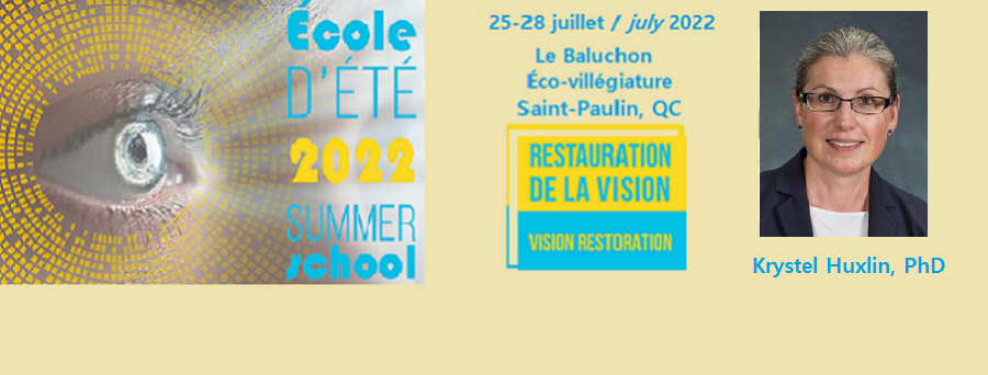
Krystel Huxlin, PhD
James V. Aquavella Professor of Ophthalmology
Associate Chair for Research | David & Ilene Flaum Eye Institute
Associate Director | Center for Visual Science
URMC Ombudsperson
University of Rochester Medical Center (URMC)
601 Elmwood Ave Box 314 | Rochester, NY 14642 USA
Ph (585) 275-5495 | Fax (585) 276-2432 | email: khuxlin@ur.rochester.edu
***
Biographie/Biography
Dr. Krystel Huxlin is the James V. Aquavella Professor and Associate Chair for Research in the Department of Ophthalmology and Flaum Eye Institute at the University of Rochester. She also serves as the Associate Director of the Center for Visual Science and co-Director of its Training program. She holds secondary appointments in the Institute of Optics, the Departments of Neuroscience and Brain & Cognitive Sciences at the University of Rochester. Dr. Huxlin earned her bachelor’s (1991) and doctorate (1994) degrees in Neuroscience at the University of Sydney, Australia, before joining the Ophthalmology faculty at the University of Rochester (1999). Her work seeks to understand how visual functions can be restored after damage to the adult visual system. She holds 9 patents and divides her attention between developing perceptual training strategies to induce vision restoration in stroke patients, and manipulating molecular substrates of corneal wound healing to prevent and treat scarring, and to restore optical quality following insults to the ocular surface. She is also part of the team that developed LIRIC, a novel, non-surgical paradigm for laser refractive error correction. She was the inaugural President of the Rochester SFN Chapter, is a reviewing editor for eLife and was just elected to the Board of Directors of VSS.
***
Résumé/Abstract
Title: A Vision For The Future: How To Recruit Neural Plasticity For Sight Restoration After Stroke
In humans, damage to the primary visual cortex (V1) causes a large loss of vision referred to as cortically-induced blindness (CB). Currently, this affects up to half a million new cases per year in the US from stroke alone, with rates rising worldwide. A major barrier these patients encounter is that in contrast with early-onset physical therapies prescribed to rehabilitate motor cortex damage, there are no accepted vision restoration therapies for CB. Over the last 15 years, the assumption that damaged, adult visual systems cannot recover functionally has been challenged by research in both humans and animal models. Human studies, which have been largely restricted to chronic CB patients whose deficits are deemed stable and permanent, point to one method consistently able to recover vision after V1 damage: visual training to detect or discriminate stimuli in the blind field. Our group was responsible for key developments in this approach, leading to mechanistic insights, but also uncovering key barriers to implementation. Among them is the fact that recovery in chronic CB requires months of daily training and the vision restored is low-contrast, coarse and restricted to the blind field perimeter, limiting its usefulness in everyday life. Evidence suggests that some of these limitations may arise because some chronic patients lose neurons that contribute to vision fundamentals not only in V1, but also – through trans-synaptic, retrograde degeneration – in the thalamus and retina. In contrast, there is little evidence of such degeneration in subacute CB patients (<3 months post-stroke). Moreover, when trained, subacute patients recover the same discrimination abilities as chronic patients, but do so 6 times faster, and with recovery extending deeper into their blind field. These data form a strong premise for refocusing investigative efforts on the substantial differences in plastic potential between subacute and chronic stroke-affected visual systems, and on defining how they can best be exploited to maximize visual restoration in CB.
***


