

Immunohistochemical evidence of synaptic retraction, cytoarchitectural remodeling, and cell death in the inner retina of the rat model of oygen-induced retinopathy (OIR)
Dorfman AL, Cuenca N, Pinilla I, Chemtob S, Lachapelle P. Invest Ophthalmol Vis Sci. 2011, 52(3):1693-708.
Impact scientifique: La rétinopathie du prématuré est l’une des principales causes de perte de vision chez l’enfant et surviens après une vascularisation anarchique de la rétine. Cette maladie est multifactorielle, et plusieurs éléments encore mal caractérisés empêchent de bien comprendre son développement. Cet article propose un nouveau mécanisme de mort cellulaire et de rétraction synaptique qui serait à l’origine des dommages causés par l’hyperoxie dans les rétinopathies du prématuré. De plus, ces travaux suggèrent fortement que des facteurs génétiques peuvent être à l’origine d’une plus grande susceptibilité au stress oxydatif entre individus. Ces mécanismes novateurs vont améliorer notre compréhension du développement de cette maladie et pourraient mener à de nouvelles pistes thérapeutiques.
Contribution du réseau: Ces travaux sont une collaboration entre plusieurs membres du RRSV et des chercheurs internationaux (Cuenca N, Pinilla I, Espagne).
* * *
Résumé original
Purpose: Postnatal exposure to hyperoxia destroys the plexiform layers of the neonatal rat retina, resulting in significant electroretinographic anomalies. The purpose of this study was to identify the mechanisms at the origin of this loss. Methods: Sprague-Dawley (SD) and Long Evans (LE) rats were exposed to hyperoxia from birth to postnatal day (P) 6 or P14 and from P6 to P14, after which rats were euthanatized at P6, P14, or P60. Results: At P60, synaptophysin staining confirmed the lack of functional synaptic terminals in SD (outer plexiform layer [OPL]) and LE (OPL and inner plexiform layer [IPL]) rats. Uneven staining of ON-bipolar cell terminals with mGluR6 suggests that their loss could play a role in OPL thinning. Protein kinase C(PKC)-α and recoverin (rod and cone ON-bipolar cells, respectively) showed a lack of dendritic terminals in the OPL with disorganized axonal projections in the IPL. Although photoreceptor nuclei appeared intact, a decrease in bassoon staining (synaptic ribbon terminals) suggests limited communication to the inner retina. Findings were significantly more pronounced in LE rats. An increase in TUNEL-positive cells was observed in LE (inner nuclear layer [INL] and outer nuclear layer [ONL]) and SD (INL) rats after P0 to P14 exposure (425.3%, 102.2%, and 146.3% greater than control, respectively [P < 0.05]). Conclusions: Results suggest that cell death and synaptic retraction are at the root of OPL thinning. Increased TUNEL-positive cells in the INL confirm that cells die, at least in part, because of apoptosis. These findings propose a previously undescribed mechanism of cell death and synaptic retraction that are likely at the origin of the functional consequences of hyperoxia.
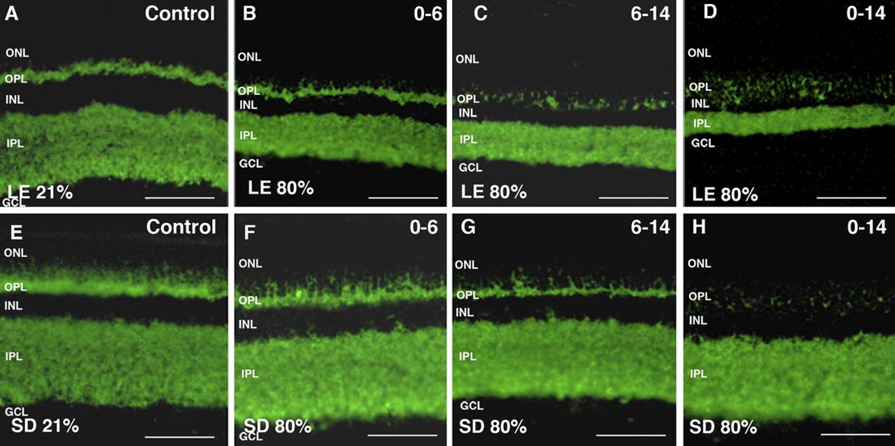
Marquage immunofluorescent indiquant une baisse des cellules neuronales dans la rétine de deux espèces de rats (Long Evans A-D et Sprague Dawley E-H) durant des expositions prolongées à l’hyperoxie.